Capnography for ROSC patients

There was a case recently where a paramedic was working a code on a patient in PEA. During the code, the EtCO2 of the patient rose from 12mm to 27. The medic correctly assumed that his patient was a ROSC patient. He stated so to the ED staff when he turned the patient over to them. The doctor ignored the medic, and stated that the increased EtCO2 meant nothing, and declared the patient dead. Some time later, when the staff went in to prepare the patient’s body for delivery to the morgue, they noticed the patient trying to breathe. The code was resumed, with the result that the patient finally expired several days later. Lawsuits are pending.

The evidence is overwhelming, capnography is a reliable predictor of ROSC in an arrest patient. Here are some studies to prove my point:

Analysis of the Efficacy of Waveform Capnography Monitoring Using Bag-Valve-Mask Ventilation. Wales, RA. Society for Technology in Anesthesia 2011.
  End Tidal Carbon Dioxide Levels Predict Cardiac Arrest. Manyam H. Society for Technology in Anesthesia 2011.
End Tidal Carbon Dioxide Predicts Cardiac Arrest. Manyam H, Thiagarajah P, Patel G, French R, Chaluvadi S, Balaan M. American Heart Association  Resuscitation Science Symposium 
Analysis of the Efficacy of Waveform Capnography Monitoring Using Bag-Valve-Mask Ventilation. Wales RA, Dyott W. Prehospital Emergency Care (NAEMSP). January 2010; Volume 14, (Suppl 1), 48-49.    
  Capnography as a Survival Predictor in Cardiopulmonary Resuscitation.Farinha LF. Resuscitation, Volume 81, Issue 2, Supplement 1, December 2010, Page S57.Sleep and Pulmonary Rehab
Partial Pressure of End-tidal Carbon Dioxide – Reliable Criteria for Termination of Non-traumatic Cardiac Arrest Resuscitative Efforts in the Field. Grmec et al. Resuscitation, Volume 81, Issue 2, Supplement 1, December 2010, Page S26.
Capnography during CPR in SUMMA 112: Preliminary Study. Diez-Picazo LD, et al. Congreso Nacional SEMES 2009. June 2009.
   

Drug lethality

Of the drugs that Americans routinely abuse, many of them are quite toxic. Take a look at this:

The median lethal doses for different substances in a 100kg male:

nicotine 5 g
Cocaine 9.5 g
aspirin 20 g
THC (main ingredient of marijuana) 127g
alcohol: 180 g
caffeine 192 g

acetaminophen (Tylenol): 200 g
table salt: 300 g

Sucrose (table sugar): 2900 g

The issue here is not so much the total lethal dose, but how close you must be to the lethal dose in order to get the effect you are looking for. The largest cluster of substances has a lethal dose that is 10 to 20 times the effective dose: These include cocaine, MDMA (often called “ecstasy”) and alcohol. A less toxic group of substances, requiring 20 to 80 times the effective dose to cause death, include Rohypnol (flunitrazepam or “roofies”) and mescaline (peyote cactus). The least physiologically toxic substances, those requiring 100 to 1,000 times the effective dose to cause death, include psilocybin mushrooms and marijuana, when ingested. I’ve found no published cases in the that document deaths from smoked marijuana, so the actual lethal dose is a mystery. My guess is that smoking marijuana is more risky than eating it, but still safer than alcohol.

Alcohol ranks at the dangerous end of the toxicity spectrum. So despite the fact that about 75 percent of all adults in the United States enjoy an occasional drink, it must be remembered that alcohol is toxic. This makes me wonder why we spend so much money on the drug war. Nevermind, the answer is profit.

Using Capnography in ACLS

You have a patient in SVT. (for more on SVT, see this post) How do you decide whether to cardiovert or use drugs? The ACLS classes recommend that you look for signs of poor perfusion, such as altered mental status or a systolic blood pressure below 90mmHg. The problem with waiting for these signs is that they are relatively late signs of cardiogenic shock.

A person with altered mental status or an SBP below 90mmHg is already decompensating, and is into the third (or possibly fourth) stage of shock. The blood pressure and mental status changes indicate that the brain and other vital organs are not being oxygenated, and as we all know, this leads to acidosis and cell death.

If only there were a way that we could determine that our patient’s SVT is reducing cardiac output due to Starling’s Law. Wish no more, here is your answer:

As cardiac output decreases, so does venous return to the lungs and heart. This lower blood flow results in less CO2 making it to the lungs to be exhaled. If your SVT patient is placed on capnography, and the CO2 is less than 37mmHg, you need to start thinking that your patient is having a bit of trouble perfusing, most likely to reduced cardiac output. This is the beginning of our patient’s journey to decompensation city. It is time to begin leaning towards cardioverting our patient before we reach the point where our patient has deteriorated so severely that he has begun to lose brain cells.

Don’t fall behind the curve. Be proactive, and you will do yourself and your patients a true service by catching problems earlier rather than later.

This happened to me

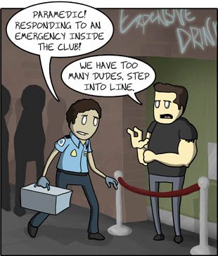
I know the cartoon is funny, but something similar happened to me about five years ago. We were dispatched to a reported drug overdose at one of our local Hispanic night clubs. (For those who are unaware, more than 45% of the local population speaks Spanish as a primary language. There is no shortage of Spanish dance clubs around Central Florida.) A young lady had called 911 to report that she and her friend were in the restroom of the club, and her friend had overdosed on an unknown drug. When we arrived on scene in the rig with all of the emergency lights flashing, I got out and went to enter the club.

The bouncer at the door told me that I had to pay the cover charge in order to enter. Thinking that he was kidding, I laughed. He blocked the door. Pointing to my uniform, I told him that I was there on official business and brushed past him. No sooner did I enter the restroom, when I was jumped by four bouncers who carried me out through the club, with the patrons of the club shouting at me, “Get out Gringo,” and yelling Spanish obscenities. The bouncers threw me out the door, and told me that whites were not allowed inside.

I called for Police backup, and got no fewer than 15 Deputies. We wound up treating our patient, and the Manager (and his bouncers) got a lecture from the cops. No arrests were made.

SVT revisited

What is Supra Ventricular Tachycardia? I think that I did a good job explaining it here. The problem that brings this up again is yet another nurse who thinks she needs to school the dumb paramedics.

I was recently training a new paramedic, and I showed him a picture of a Sinus Rhythm at a rate of 180 beats per minute. I told him that he was, for the purpose of this discussion, working at the first aid tent of a marathon, and that this was the presenting rhythm of a marathon runner who was complaining of dizziness. I use this scenario often as a training aid, and this medic, as new medics invariably do, identified the rhythm as PSVT and stated that it should be treated with 6mg of Adenosine. I asked why, and he told me that PSVT is any rhythm that has an origin above the ventricles, and a rate of over 150 beats per minute.

I pointed out to him that while SVT is technically any tachycardia that occurs above the ventricles in a purely literal sense, it is important for clinicians to recognize that what we refer to as PSVT is a dysrhythmia, and that there is not any set heart rate that separates Sinus Tachy from PSVT. The only way to tell the difference is to do a good patient assessment.

A nearby RN overheard, and attempted to tell me why I was wrong, and that 150 bpm is the standard for defining SVT.

I asked her why she felt I was incorrect in saying that the aforementioned marathon runner is probably in Sinus Tachy. This rhythm is a response to the normal metabolic demands of the runner’s body. A person taking a stress test has similar responses. (After all, the target heart rate for a stress test is usually over 150 beats per minute)

Instead of answering, the RN tried to tell me that the AHA defines SVT as all tachycardias that originate above the ventricles and have a rate over 150. Sigh. Where does the AHA say that?

The Big Red Taxi

Ask anyone who works in EMS, and they can tell you a hundred stories of people who abuse the EMS system. I have seen my share, and the stories have infinite variety:

The thermostat:
It was 2 o’clock in the morning when we went to this woman’s house for a complaint of “difficulty breathing.” When we got there, the “patient” didn’t want to go to the hospital, she just wanted help. Having just moved into her house, she didn’t know how to program her electronic thermostat and wanted us to show her how. It wsa 82 degrees in her house, nowhere near being a medical emergency.

Incarceritis:
People fake seizures, unconsciousness, chest pains, you name it, in the belief that they will not be arrested if they go to the hospital. The sad fact is that they are sometimes right. The officer sometimes doesn’t want to sit around the hospital for several hours, waiting for his prisoner to be discharged for minor misdemeanors. Felony arrests? Forget it, you are going to jail as soon as the hospital is done with you.

A variation of this was one morning’s call:
A man was required to be in court at 8 o’clock in the morning for a child support hearing. He walked from his home towards the courthouse 10 miles away. (Why he didn’t take the bus is a mystery.) He made it about 3 miles, and then called 911 with reported shortness of breath and chest pain at about 10 after 7. When we arrived, he wanted to go to the hospital that, coincidentally, was two blocks from the courthouse. When we arrived and asked him what was going on, his first words were, “I am trying to get to court because my wife…”

We all knew that this was a fake call intended to get him to the hospital closest to the courthouse, so that he could walk out and be in court faster. Since there is no penalty for failure to pay for the ambulance ride OR the ER bill, this is the equivalent of a free taxi. We decided that the best thing for this patient was to go to a different hospital. Why? Well, if he really was having chest pain, the patient would be less likely to walk out of that one, opting instead to use the ER bill to show the court why he had a medical reason for missing court. IF he wasn’t really having chest pains, then he didn’t need to go anyhow. Besides, to reach the hospital he wanted, we would have had to pass another, closer one, and there was no medical reason to pass a perfectly good hospital to go to the other one.

Lest anyone think that we were being mean or lazy, he still got a complete workup. His vitals were: SaO2 98% on room air, 100% on 2 lpm of O2. HR 82, RR24, BP 142/94. Monitor showed SR, and 12 lead showed nothing important. He was hot and sweaty, but that is unsurprising considering that he was about 60 pounds overweight, and it was 82 degrees with 80% humidity. He still got 325 mg of aspirin, NTG spray x2, and transport to the closest hospital (rather than the one he wanted)

People abuse the system every day. This is why universal, “free” health care will never work.

A lesson in cardiology

This post arises from a call I ran yesterday with a relatively new medic. Those of you in EMS know that new medics are pretty timid, and tend to have problems with calls that require them to think outside of the box we call protocol. This was one of those times that it is obvious that we need to do so. I hope any new medics that read this will learn an important lesson.

Our patient was a 58 year old man who was working at some light gardening when he began having pain between his shoulder blades, and began loudly burping, which relieved the pain. He went inside and took some gas medicine. His obviously intelligent wife called 911 and got him to take 2 baby aspirin.

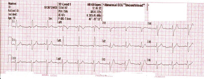
When we arrived, we found him seated on his couch, he was cool and covered in sweat. He states he has a history of hypertension, for which he takes no medicine. He states that his doctor feels like his blood sugar is too high, and wanted him tested for diabetes, but that was 6 months ago, and he hasn’t been back since. When we stand him up to plpace him on the stretcher, his pulse becomes irregular. His vitals are: P- 88, BP 138/86, RR 20. In the truck, we start an IV, and find him to be in a normal sinus rhythm, his SaO2 is 99% on room air, blood sugar is 170. His 12 lead EKG is as follows:

(Click for a larger picture)

There are a few things that jump out at you here.

  1. The 12mm height of the QRS in lead aVL indicates “voltage criteria” for left ventricular hypertrophy. This is likely caused by his history of uncontrolled high blood pressure.
  2. The length of his QRS (0.122 sec) could be considered a left bundle branch block, but I think this is probably due to the QRS widening caused by the LVH, since the EKG doesn’t have that “LBBB look” to it.
  3. The poor R wave progression in leads V1 through V6
  4. The ST segment depression in Leads V3 and V4

I pointed out to New Medic Partner (NMP) that nitroglycerin was probably a good idea here. NMP didn’t want to give it because the patient “didn’t have chest pain.”

Sigh. This is one of my pet peeves. This man has risk factors- possible untreated diabetes AND high blood pressure. He is complaining of anginal equivalents- indigestion, and back pain. His irregular pulse on standing MAY be PVC’s, and his 12 lead confirms a cardiac event.

I finally prevailed, and we alerted the hospital. The doctor sent him to the cath lab, and it turns out that he had a complete blockage of the distal end of the left anterior descending coronary artery. He was having a hearta ttack, but we caught it early, and he is now recovering.

Please, medics. Learn how to read the signs your patient is giving you, and learn to read and interpret the 12 lead EKG. Cardiology, diabetes, and drug overdoses are the three areas where medics save the most lives. Be the best you can be at this, lives depend on it.

Half Assed Medic

WARNING: Medical terminology to follow, but I will try and keep it to a minimum.

I was the first medic on scene to a 54 year old male, whose chief complaint was that he nearly passed out while he was lifting a heavy object from the back of his minivan. He had a History of insulin dependent diabetes, a heart bypass, high cholesterol, and high blood pressure. His vitals were as follows: HR 72, RR 20, BP 136/72. He takes lopressor, insulin, lipitor, and aspirin. His 12 lead showed nothing acute, except LVH. I was in the middle of finishing my assessment when the transport unit arrived.

The medic on that truck told the patient that the near syncope was probably due to stimulation of the vagus nerve that lifting the box caused. I pointed out to the patient that while the other medic was probably correct, due to his extensive history, it would probably be best to take him in, just to be sure. You see, diabetics frequently do not have the classic symptoms of a heart attack, and often the first sign that a diabetic has of a serious heart attack is fainting, nausea, or shortness of breath, and not chest pain. The patient agreed with me, and decided to go to the hospital.

Apparently, that angered the other medic, who had been hoping to talk the patient out of going to the hospital. After he dropped the patient off at the hospital, he decided to come talk to me about “taking over his patient.” I pointed out to him that he is a new medic (less than a year on the street) and that patient refusals are not there for his personal comfort- paramedic inconvenience is not a reason to avoid transport.

If there is any piece of advice I can give you new medics out there, it is this:

If you are ever undecided as to the proper course of action, whichever option it is that causes you the most work is usually the correct one. Don’t ever forget that we are the patient’s advocate, and all of our decisions need to be in the best interests of the patient, not ourselves.