Tourniquet Conversion

Scientific knowledge evolves. As mankind discovers more information, the view of our world and how it works changes as well. The use of tourniquets, which I have discussed at length a couple of times on this very blog, are a great example. We were once taught that, once a tourniquet goes on, it stays on unless the patient is in the room with a trauma surgeon. That turns out to be only partially correct.

At the end of the day, trauma is a surgical emergency. What a trauma patient needs is access to that trauma surgeon. Getting a patient to the hospital within a rapid timeframe, often referred to as “the golden hour” greatly increases the patient’s chances of survival. Remember that serious trauma is first and foremost a surgical emergency. Trauma patients don’t need a tricked out first aid kit- they need a trauma surgeon. All they need you to do in the field is keep them alive and prevent them from furthering their injury until they can get on the operating table.

For that reason, tourniquet training for most Americans has followed simple, strict guidance: apply one high and tight to limbs to stop life-threatening bleeding, and leave removal to advanced medical care personnel. That is no longer the case.

So what changed? The war in Ukraine. In the US, and in US controlled battlefields, people who receive gunshot wounds are one helicopter ride away from a trauma surgeon, and the vast majority of the patients make it to the surgeon’s operating table within that hour and have good results. In Ukraine, the nature of the war there is such that almost no one gets a helicopter ride, and with killer drones circling everywhere, very few of those injured are transported by ground in anything resembling even the “golden day or two” and, if they are wearing a tourniquet, wind up wearing it for days. Casualty evacuation in Ukraine often exceeds 6 hours, and the liberal use of limb tourniquets may have increased morbidity.

That sets up a whole list of new problems.

Leaving a tourniquet on for hours or even days causes other issues. Once a tourniquet is in place, it stops blood from flowing to the affected limb. Once deprived of their blood supply, those cells begin taking measures to protect themselves, and after a period of time, those cells die. As they die, they release their contents into the surrounding tissue. When the tourniquet is removed, those contents make it to the kidneys and clog them up with all of that debris. The condition is called rhabdomyolysis. The longer a limb tourniquet is in place, the higher the risk of compartment syndrome, vascular thrombosis, rhabdomyolysis, and irreversible myonecrosis resulting in major tissue loss and often necessitating limb amputation, kidney failure, and death.

If bleeding is controlled with a tourniquet, attempts to remove the tourniquet as early as possible to avoid the negative consequences are essential. However, if the removal is not done correctly, there is a risk the bleeding that the tourniquet was supposed to stop will also kill the patient. The act of removing a tourniquet (called ‘tourniquet conversion’) is now changing the training being put out to US special forces troops, and is especially important to preppers here in the US.

What this means for you

In the US, we are usually a quick helicopter ride away from a trauma surgeon, but that may not always be the case. In a disaster or TEOTWAWKI situation, those helicopters aren’t coming. At the same time, a tourniquet left on for more than 2 hours can itself be a hazard. Before attempting this, please review my post on treating gunshot wounds that you can find here.

Important: Never attempt tourniquet conversion if the patient is in shock, the wound cannot be continuously monitored for re-bleeding, or if the tourniquet is applied above a traumatic amputation.

Shock being defined here as the loss of consciousness, or a blood pressure less than 90/xx.

The Conversion Steps (The “Plus-1” Method)

  1. Prepare: Place a second, loose tourniquet on the bare limb just above the first one, but do not tighten it yet.
  2. Pack: Firmly pack the wound cavity with hemostatic gauze (if available) or standard rolled gauze, maintaining direct digital pressure for at least 3 minutes.
  3. Dress: Apply a pressure dressing securely over the packed wound.
  4. Release: Slowly loosen the original tourniquet (over at least 1 minute) while meticulously observing the wound for bleeding.
  5. Monitor: If bleeding is controlled, leave the primary tourniquet in place but fully loosened in case bleeding recurs.

Optimal conversion should be attempted within 2 hours of the initial application when conditions are safe and stable. If bleeding restarts upon release, re-tighten the primary tourniquet or utilize the newly placed backup tourniquet.

For more information, see the article Tactical Medicine Tourniquet Conversion or consult the sources below:

Sources:

Misuse of Tourniquets in Ukraine may be Costing More Lives and Limbs Than They Save.

Rethinking limb tourniquet conversion in the prehospital environment (pdf warning)

Tactical Combat Casualty Care skill card: Tourniquet Conversion (pdf warning)

Tourniquet Conversion: A Recommended Approach in the Prolonged Field Care Setting

Tourniquet Conversion: The Critical Skill Nobody Teaches After You Stop the Bleeding (2026)

Tough Guy

This is a story of a patient from a year or so ago, while I still worked for my last employer.

A patient comes in because he has had increasing shortness of breath for about two weeks. He was walking down a small hill from where he parked his motorcycle and fell, tumbling down the small incline. He fell about 10 feet or so, he says. His vitals look fine. He is a bit of an overweight guy, typical 60-something man trying to recapture his youth by riding a Harley.

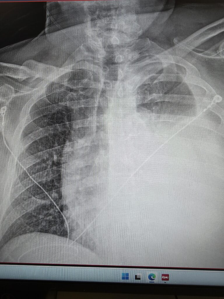
So I ordered a chest x-ray, started an IV, and did his intake paperwork. No doctor is signed on to his case yet. If the x-ray shows anything significant, the technician who takes it will normally give me a heads up. He didn’t in this case. The image of the x-ray came up on my computer, I took one look at it and immediately flagged down the first passing doctor and said, “Hey, I know that you’re busy and this isn’t your patient, but you need to see this now.” Here is what it looked like:

In case you don’t know what you are looking at, the dark section on the left is a relatively normal looking lung. The heart and trachea are supposed to be on the right side of the image and are being pushed into the other side by the large amount of blood that is collapsing his left lung (which appears on the right in this image). If you look closely at the film, you can see all of the structures that are supposed to be midline are being pushed over. This is called a hemothorax, and is a life-threatening medical emergency where a massive volume of blood rapidly accumulates in the pleural space (the area between the chest wall and the lung). This buildup compresses the lung and puts dangerous pressure on the main vein bringing blood to the heart (the vena cava) and the heart, leading to cardiovascular collapse, severe respiratory distress, and shock.

The doctor took one look at this and said “Holy shit! I’m signing up for him. Get me set up for a chest tube and some conscious sedation. Call respiratory and let’s get ready to send him to a trauma center.”

The patient had a rather chubby neck with a beard so it wasn’t readily apparent, but if you put the finger of one hand on his Adam’s apple, and a finger from the other hand in his sternal notch, you could see that his windpipe was deviating to the patient’s right. He was a good sport and didn’t even mind that I brought a couple of new nurses into the room to see what a tension hemothorax looked like. Of course, he had no lung sounds on the left, and his heart tones were distinctly muffled. His pulse pressure was a bit narrow.

There were not any other nurses or respiratory technicians available to help in time, so I grabbed a nursing assistant and the three of us (doctor, myself, and aide) rapidly initiated conscious sedation and inserted a chest tube. That’s a handful for one nurse and a doctor to handle (the nurses aid is pretty much there to hold this, and hand me that and isn’t much of a help)- I had to administer sedation, monitor and maintain his airway and breathing, and chart everything. For one nurse to do all of that without help is a major safety issue, and is one of the (many) reasons why I don’t work for that hospital any longer. That place is just understaffed to the point of compromising patient safety.

Once we got the tube in place, we sent him for a CT scan, and it turns out he had 4 ribs broken in two places- a classic flail chest. If you put your hands on his rib cage, you could feel the paradoxical motion of the chest wall. This is incredible, considering that he walked in to the ED and had been walking around like this for two weeks. The video below shows you what paradoxical motion looks like, but my patient’s wasn’t quite as pronounced as the video (and was located under his left armpit).

Anyhow, I pulled about 2 liters of blood from his chest cavity before we crimped off the tube because we didn’t want him losing too much blood. A helicopter came and took him to a trauma center, and the trauma surgeon was still pissed because we took out so much blood.

The patient made a full recovery.

Old School

A few of my readers have commented on how they enjoy practice pearls, so I thought I would go ahead and share a simple one. This particular pearl is not just for medical people, it will work quite well for those of you who are not in the medical profession. In the medical profession, a lot of time is spent training people in the latest, greatest technology- the newest medication, the latest technique, the wow factor, but sometimes, it is the old school method that works best. I recently had the chance to teach a young doctor this exact lesson.

Lacerations

We had a woman that entered the emergency room who had cut off the last quarter inch or so of the tip of her finger with a pair of scissors while attempting to cut open an Amazon package, of all things. In so doing, she had also managed to nick the artery, and blood was pulsing out of the end of her finger. She had the presence of mind to bring in what she thought was the tip of the finger (what she brought in wasn’t the finger) and she was attempting to control the bleeding herself, but wasn’t succeeding.

The doctor looked at it and suggested we perform a digital block and attempt to stitch off the artery and suture the wound closed. That was going to be a bear to do. Instead, I suggested that we try something a bit more old school. I took a large emesis basin, filled it with ice, a little water water, and a bottle of Providone. I told the woman to place her finger in the basin and keep it there until the cold became painful. This concoction does three things:

  • The ice numbs the area
  • the Providone cleans the wound (there is a risk of infection if you don’t)
  • the ice also constricts blood vessels and is great at controlling bleeding

She spent about 15 minutes with her hand in that basin, and this stopped about 80% of the bleeding. I pulled her hand out, cleaned the finger with a couple of gauze pads (4×4), then had her hold one against the tip of her finger using her thumb. After about 5 minutes of that, we still had some bleeding, so I soaked a gauze pad with tranexamic acid (TXA) and placed it against the wound. That stopped the rest of the bleeding. I dressed the wound, and we sent her home.

Esophageal Varices

Just as you can get varicose veins in your legs, you can get them in your esophagus as a result of cirrhosis of the liver and the resulting portal hypertension. I was working in the ED one night when a patient began vomiting large amounts of blood. I’m not talking about what most people would think are large amounts of blood, but what a nurse who works in the ED thinks are large amounts of blood. It looked like this:

We were under the gun: if this bleeding didn’t stop, we were looking at a dead patient. I inserted an NG tube to suction out the blood, and the doctor and I came up with a plan. Using a piston syringe, I would push about 100 ml of ice water into the tube, let it sit for about 30 seconds, then suction it back out. I repeated this about 5 or 6 times, and each time, the amount of blood that came out with it was less. I was forced to stop after that 5th or 6th time, because the cold must have been irritating to her heart, as evidenced by the fact that she began having short runs of ventricular tachycardia. For that reason, anyone trying this, I would recommend placing the patient on a cardiac monitor and keeping a close eye on on their heart rhythm while doing this.

Ice- it’s quite useful in emergency medicine, but it isn’t used much any more in emergency medicine because it doesn’t have the sexy feel of the latest, greatest advances in medical technology, but it is still damned effective. Sometimes old school is still the best way to go.